Fungal Skin Discoloration Self-Checker
Answer the questions below to get a preliminary assessment of possible fungal skin discoloration.
Do you notice any of these symptoms?
Spotting a change in skin tone can be unsettling, especially when you’re not sure whether it’s harmless or something that needs treatment. Fungal skin discoloration often begins subtly - a faint patch here, a slightly lighter spot there - and it’s easy to dismiss until it spreads. This guide will walk you through the tell‑tale clues, how to tell it apart from other skin issues, and what steps to take before the problem gets bigger.
TL;DR - Quick Takeaways
- Look for uneven color patches that appear suddenly, especially on the chest, back, or shoulders.
- Early signs include mild itching, flaky texture, and color that’s lighter (hypopigmented) or darker (hyperpigmented) than surrounding skin.
- Common triggers: heat, sweat, oily skin, and sun exposure.
- Self‑check: wash the area, let it dry, and observe if the patch changes after a few hours.
- See a dermatologist if the patch persists beyond two weeks or spreads rapidly.
What Is Fungal Skin Discoloration?
When we talk about fungal skin discoloration is a change in skin color caused by an overgrowth of certain fungi on the surface of the skin, we’re usually dealing with a condition called Tinea versicolor (pityriasis versicolor). The culprit is a type of yeast called Malassezia, which lives harmlessly on most people’s skin. When conditions such as heat, humidity, or excess oil tip the balance, this yeast multiplies and produces pigments that alter the skin’s natural shade.
Early Signs to Watch For
Fungal discoloration doesn’t usually scream “danger” right away. Instead, you’ll notice a handful of subtle cues:
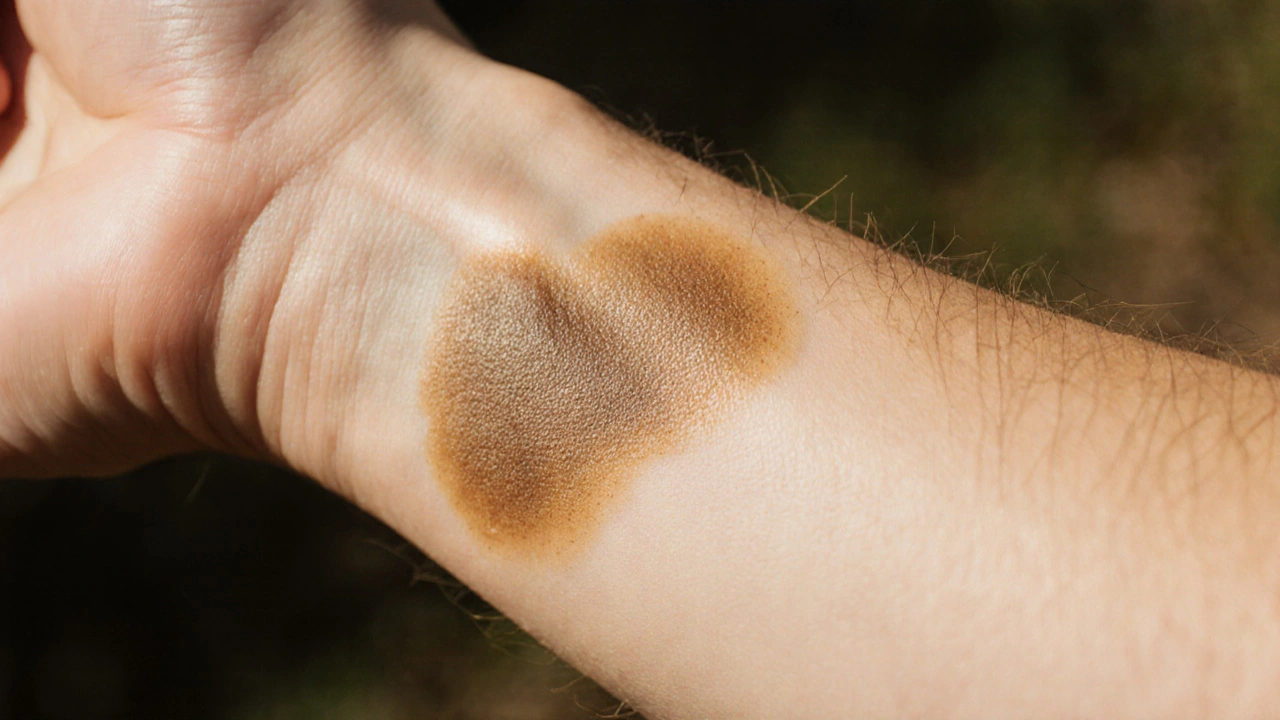
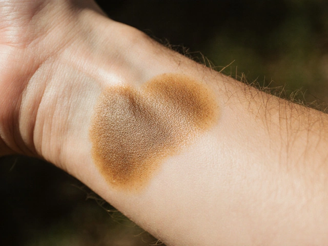
- Uneven patches - Small to medium‑sized spots (1‑5cm) that are lighter (white‑gray) or darker (brown‑tan) than the surrounding skin.
- Mild itching or tingling - Often described as a faint crawl, especially after sweating.
- Fine scaling - The patches may feel slightly rough or flaky, like after a mild sunburn.
- Color change after sun exposure - The affected area may stay lighter while the surrounding skin tans, making the contrast more obvious.
- Location patterns - Common on the chest, upper back, shoulders, and sometimes the neck or arms.
If you spot a combination of these signs, it’s worth doing a quick self‑check.
How to Differentiate From Other Skin Conditions
Not every patch of discoloration is fungal. Here’s how you can tell the difference:
| Condition | Typical Color | Texture | Location | Trigger |
|---|---|---|---|---|
| Tinea versicolor | Light‑gray, tan, or brown | Fine scaling, mild itching | Chest, back, shoulders | Heat, sweat, oily skin |
| Vitiligo | Pure white, well‑defined | Smooth, no scaling | Any area, often around eyes, mouth | Autoimmune, genetic |
| Eczema (atopic dermatitis) | Red, sometimes darkened after scratching | Very dry, thickened, itchy | Elbows, behind knees, neck | Allergens, stress |
| Psoriasis | Silver‑white plaques | Thick, silvery scales | Scalp, elbows, knees | Immune system triggers |
Notice that fungal patches tend to appear suddenly and are linked to warm, sweaty environments, whereas conditions like vitiligo develop gradually and have distinct borders.
Risk Factors and Triggers
Understanding what fuels the fungus helps you spot early warnings. The main contributors are:
- Sun exposure - UV light darkens normal skin, leaving fungal‑affected areas lighter in contrast.
- Sweat and humidity - Moisture creates a perfect breeding ground for Malassezia.
- Oily skin - Excess sebum feeds the yeast, especially on the back and chest.
- Hormonal shifts - Puberty, pregnancy, or hormonal meds can alter oil production.
- Immune suppression - Certain medications or illnesses reduce the skin’s natural defenses.
If you notice a pattern - like a new patch after a holiday beach trip - that’s a strong hint the fungus is at work.
Self‑Check: Steps to Confirm Suspicion
Before you book an appointment, try this simple at‑home test:
- Clean the area with a gentle cleanser and pat dry. Avoid harsh scrubs.
- Observe the texture - run a fingertip over the patch. Light scaling is a clue.
- Apply a piece of white paper or a cotton swab and lightly press. If the patch leaves a faint imprint, it’s likely a superficial fungal layer.
- Expose to sunlight briefly (10‑15 minutes). After a few hours, the unaffected skin will tan, making the lighter patch stand out.
- Monitor for 7‑10 days. If the discoloration persists or spreads, it’s time to see a professional.
This test isn’t a substitute for a medical diagnosis, but it helps you decide whether a doctor’s visit is warranted.
When to See a Dermatologist
Schedule a consultation if you observe any of the following:
- The patch enlarges by more than 1cm per week.
- Intense itching, burning, or pain develops.
- Discoloration appears on the face, genitals, or near the eyes.
- Over‑the‑counter creams (like topical zinc pyrithione) don’t improve the area after two weeks.
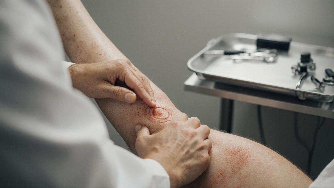
A dermatologist can perform a Wood’s lamp examination (blue‑light) - fungal patches will glow a yellow‑green hue - or take a skin scraping for lab analysis.
Prevention and Early Treatment Options
Even if you catch the problem early, treating it right away reduces the chance of recurrence.
- Topical antifungal creams - Look for active ingredients such as clotrimazole, ketoconazole, or selenium sulfide. Apply twice daily for at least two weeks, even if the patch fades sooner. \n
- Shower after sweating - Quickly rinsing off sweat and drying thoroughly keeps the yeast from proliferating.
- Use medicated shampoos on the back and chest - Even non‑hair areas benefit from a weekly wash with a selenium sulfide shampoo.
- Wear breathable fabrics - Cotton and moisture‑wicking sportswear reduce trapped heat.
- Limit oily skin products - Heavy lotions can feed the fungus.
For stubborn cases, doctors may prescribe oral antifungals like terbinafine for a short course, but these come with monitoring requirements, so they’re reserved for extensive involvement.
Quick Comparison Table: Fungal Discoloration vs. Mimicking Conditions
| Feature | Fungal Discoloration (Tinea versicolor) | Vitiligo | Eczema |
|---|---|---|---|
| Onset | Sudden, weeks | Gradual, months‑years | Intermittent flares |
| Color | Light‑gray to brown | Pure white | Red, may darken after scratching |
| Scaling | Fine, sand‑paper feel | None | Thick, dry |
| Triggers | Heat, sweat, oily skin | Autoimmune | Allergens, stress |
| Treatment | Topical/oral antifungals | Light therapy, steroids | Moisturizers, steroids |
Next Steps: Your Action Plan
Now that you know what to look for, here’s a simple checklist you can keep in your phone or on the fridge:
- Notice any new patch? Note size, color, and feeling.
- Run the self‑check steps (clean, test, sun‑expose).
- If the patch lingers >10days, try an OTC antifungal cream.
- Re‑evaluate after 2weeks - improvement? Keep using cream.
- No change or worsening? Book a dermatologist appointment.
Consistent early detection saves you time, money, and the frustration of a spreading rash.
Frequently Asked Questions
Can fungal skin discoloration turn into skin cancer?
No. Fungal discoloration is caused by yeast, not by atypical cell growth. However, any persistent skin change should be evaluated to rule out unrelated issues.
Why does the patch look lighter after a sunny day?
Sun exposure tans the healthy skin but doesn’t affect the fungus‑produced pigment, so the affected area stays its original shade, creating a stark contrast.
Is a home remedy like apple cider vinegar effective?
Anecdotal reports exist, but scientific evidence is limited. A proven OTC antifungal with clotrimazole or ketoconazole offers reliable results.
Can I wear sunscreen over a fungal patch?
Yes, sunscreen won’t harm the treatment and can reduce the visual contrast that makes the patch more noticeable. Choose a non‑greasy, fragrance‑free formula.
How long does treatment usually take?
Topical creams are applied for 2‑4 weeks, even if the spot fades earlier. Oral medication, when prescribed, often runs 2‑3 weeks.


Oliver Bishop
September 29, 2025 AT 20:59Nothing beats good skin health, folks-keep it clean!
Alissa DeRouchie
September 30, 2025 AT 15:02Honestly I think most people just ignore the signs and it’s a disaster waiting to happen, the whole “just a little patch” excuse is so overused and lazy.
Emma Howard
October 1, 2025 AT 09:06Exactly! You’re spoton! Let’s all stay proactive and use an antifungal cream early – you’ll thank yourself later!
Courtney Payton
October 2, 2025 AT 03:09I find it morally imperative to educate people about fungi, yet too many just skip the shcure and ignore the real issue.
Muthukumaran Ramalingam
October 2, 2025 AT 21:12Look man, I get that you’re trying to be all moralistic, but honestly the patch thing is just a skin thing you can wash off, dry and maybe use some over‑the‑counter stuff. It’s not rocket science. If you keep sweating and not drying properly, the fungus loves that. So yeah, just keep clean, no need to get all preachy about it.
Garrett Williams
October 3, 2025 AT 15:16Stay positive, a quick soap and a good antifungal can make a big difference.
joba alex
October 4, 2025 AT 09:19While your lay‑man advice is appreciated, let’s consider the dermatological terminology: hyperpigmented versus hypopigmented lesions, Malassezia spp. proliferation, and the role of sebaceous activity in the pathogenesis.
Rene Lacey
October 5, 2025 AT 03:22When we contemplate the subtle emergence of a discoloration, we are really observing the dialogue between our integumentary system and its microscopic inhabitants.
The fungus, chiefly Malassezia, exploits the biochemical milieu of warm, oily skin to produce pigments that alter the visual spectrum.
This process is neither instantaneous nor catastrophic, but a gradual shift that can be mapped through careful self‑examination.
The early signs-uneven hue, mild pruritus, and a faint scaling-function as physiological breadcrumbs.
By paying attention to these breadcrumbs, one can intervene before the pattern expands across larger dermal territories.
Topical azoles such as ketoconazole or clotrimazole penetrate the stratum corneum and disrupt the fungal cell membrane.
Consistent application, typically twice daily for a fortnight, ensures that residual spores are eradicated.
In addition to pharmacologic measures, environmental control plays a pivotal role; humidity, sweat, and occlusive fabrics create a micro‑ecosystem favorable to fungal growth.
Thus, wearing breathable cotton, showering promptly after exertion, and avoiding heavy lotions constitute non‑pharmacologic prophylaxis.
Should the lesions persist beyond two weeks, a Wood’s lamp examination can reveal the characteristic yellow‑green fluorescence.
This diagnostic adjunct aids clinicians in differentiating tinea versicolor from vitiligo or eczema.
Moreover, oral agents like terbinafine are reserved for extensive involvement due to systemic side‑effects.
Patients often underestimate the psychosocial impact of visible skin changes, which can affect confidence and social interaction.
Addressing the condition early not only restores the aesthetic appearance but also mitigates potential secondary infections.
In sum, a vigilant self‑check routine coupled with timely therapeutic action embodies the optimal strategy against fungal skin discoloration.
johnson mose
October 5, 2025 AT 21:26Wow, that breakdown hits the sweet spot – the science, the self‑care, and the confidence boost all wrapped in one clear guide.
Charmaine De Castro
October 6, 2025 AT 15:29Great summary! I’d add that using a non‑greasy sunscreen can help hide the contrast while you’re treating the area.
Mark Mendoza
October 7, 2025 AT 09:32Absolutely 🌞! Sunscreen won’t interfere with antifungal creams and keeps the surrounding skin from darkening 🌿.
Dan Tourangeau
October 8, 2025 AT 03:36Remember to wash the spot, dry thoroughly, and monitor for a week before seeking a dermatologist.
Bernard Valentinetti
October 8, 2025 AT 21:39Indeed; the procedural rigor of cleansing, desiccation, and temporal observation constitutes the gold standard of pre‑clinical dermatological assessment 🤓✨.
Kenneth Obukwelu
October 9, 2025 AT 15:42From a cultural perspective, many societies have traditional remedies for skin balance, yet modern antifungals provide a scientifically proven path.
Josephine hellen
October 10, 2025 AT 09:46It’s fascinating how age‑old practices like tea tree oil or sandalwood paste echo today’s emphasis on keeping the skin environment hostile to fungal overgrowth; however, clinical trials show that standardized ketoconazole ointments deliver a more reliable eradication, so blending tradition with evidence‑based medicine can give the best of both worlds, especially for those who cherish their heritage while seeking effective results.
Ria M
October 11, 2025 AT 03:49In the grand theater of the epidermis, the fungal actor performs a subtle yet pivotal role, and recognizing its cues is an act of enlightened self‑care.
Michelle Tran
October 11, 2025 AT 21:52True 😅 – just wash and move on!